At BSMS, the anatomy department have incorporated new digital teaching resources to aid teaching during the Covid-19 pandemic. This significant increase in digital resources will go on to benefit students for many years to come.
Live streaming
It was important to us that we still gave students exposure to human cadaveric material, albeit virtually. One of the main methods we used to achieve this was live streaming. At the beginning of the pandemic, anatomy at BSMS invested in new streaming camera equipment. This allowed us to stream straight from the dissection room to hundreds of students at once.
Over the past year we have live streamed numerous sessions to replace the face-to-face practical sessions that would normally run. These sessions normally fall into one of three types: dissection, prosection or living anatomy and ultrasound. Dissection sessions involve an element of active dissection, where students are expected to reveal structures in their groups. Prosection sessions use pre-dissected specimens with all of the structures that students need to study already revealed.
Finally, living anatomy and ultrasound sessions do not use human cadaveric material but rather look at the anatomy we can identify in the living, using palpation (feeling) of structures and ultrasound imaging. At certain points during the past year, we have delivered all three types of practical teaching using live streaming.
Live streaming will continue to be used in anatomy at BSMS. Practical face to face sessions will resume for the majority of sessions but will be supplemented by live streaming for certain elements. In addition to its uses in undergraduate medical education, live streaming can also be used to deliver surgical training to clinicians across the world.
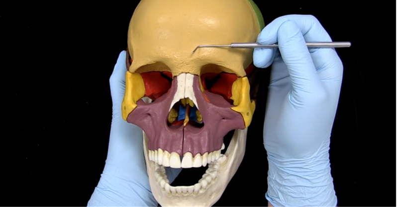
Example of view from the cadaveric videos (note: this is not a cadaveric specimen)
Anatomy learning interface
The anatomy learning interface has undergone significant development in response to the Covid-19 pandemic. The anatomy learning interface is an application that allows us to upload images and videos containing human cadaveric material in a secure manner. Students can then access this content using any handheld device. This secure platform is important due to the sensitive nature of the material, we work with.
Lots of new resources were added to the existing content on the anatomy learning interface for the students at BSMS during the pandemic. The anatomy learning interface now contains a full complement of narrated videos containing cadaveric specimens, covering all areas of anatomy.
Once face-to-face teaching has resumed, these videos can be used to supplement practical sessions. Students will be able to watch the videos before entering the dissection room as a way of preparing, or after they have left the dissection room as a way of recapping everything they covered. Students are not allowed to capture any images or bring anything into the dissection room (including materials to make notes with). These videos will therefore provide an excellent opportunity to review the content outside of the dissection room.
Students can also access images and descriptions of potted specimens through the anatomy learning interface. In the dissection room we have a whole range of historic potted specimens. Normally relevant potted specimens are brought out for students to view during their practical sessions in the dissection room. However, it is now possible for students to also access images and descriptions of these potted specimens from home through the interface.
Complete Anatomy
BSMS Anatomy also reviewed their offering of 3D virtual applications and we now have a licence for complete anatomy. Complete anatomy is a 3D virtual anatomical atlas that includes hundreds of videos, models and screens that can be used to aid understanding. Often lecturers will use complete anatomy to explain difficult concepts during a lecture. With complete anatomy we can now save all of the screens we use during lectures and provide them to students so that they can explore and manipulate them in their own time. This has been a huge benefit while delivering online teaching and we will continue to integrate it into our teaching in years to come.
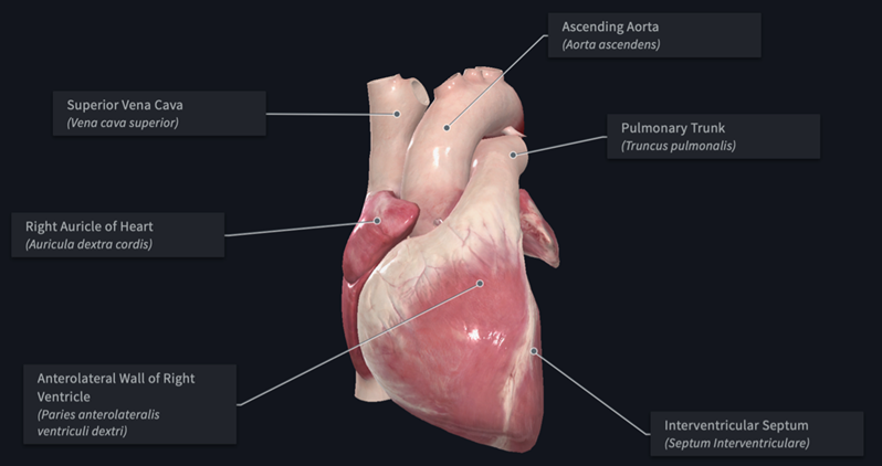
Example of complete anatomy
Take a virtual tour of our anatomy suite here >