Skip to main contentSkip to footer
Hands on an iPad featuring an anatomical image
Brighton & Sussex Medical School

TEL Case studies

BSMS > About BSMS > TEL > TEL Case Studies

TEL Case Studies

Here, we present examples of practice from teaching staff across faculty in collaboration with learning technologists. The case studies will report on the impact of the implementation of TEL across the curriculum. 

Medical history taking

Innovative Interactive Medical History video allows BSMS students to curate their own learning. 

01/09/2019 

The TEL team, directed by Physician Associate Lead Richard Howell, have created a user-controlled taking of a full medical history to provide students with an innovative reference and revision tool.  

The interactive video, created and hosted through the H5P authoring tool in Student Central, allows students to see an exemplar full medical history take with an optional audio narration to guide them through the purpose and reasoning.  

The video had a full cast consisting of a paid actor, two Doctors (Richard Kerslake, Nicola Dearnley) and a Physician Associate (Shannon Rosine). The voiceover was provided by Duncan Shrewsbury. It was filmed in the Moulsecoomb campus media studios under the guidance of Learning Technologist CJ Taylor.  

Project lead, Richard Howell, said: "We hope that this video, using three different styles, provides both a good learning and revision tool for taking a full medical history. It is intended for undergraduate medical students and PAs both for when they first embark on their courses and later for reflection and consolidation".

BSMS Learning Technologist Nicola Walters, who worked on the post-production of the project, said: "Development of interactive learning resources allows learners to curate their learning and engage with content at their own pace and convenience".  

Watch the final video on Student Central in the video resources section here > 

Anatomy interface

Digital anatomy at your fingertips: A bespoke touchscreen gateway for accessing digital anatomy resources at the dissection table. 

01/09/2019

There are a wealth of digital learning resources available to students in human anatomy, including in-house teaching material and externally sourced products. However, access to these is dispersed across interfaces which can be a barrier to an integrated teaching and learning approach in the dissection lab. 

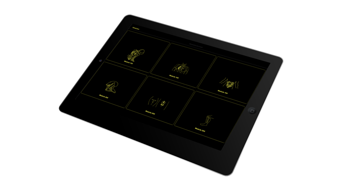
This project created a bespoke web-based interface for iPad that compiles access to multifarious digital anatomy resources into one clear and secure gateway for use at the dissection table.  

Using HTML and JavaScript, a kiosk-style menu interface was created for use on a set of laboratory-dedicated iPads. Through this gateway, students access the diverse collection of learning resources during the dissection exercises, including the PDF dissection workbook, lecture materials on the VLE, video explanations, and full e-books. 

Additionally, Brighton and Sussex Medical School has a large collection of pathology specimen pots which are labelled with a QR barcode, which when viewed with the iPad camera, provide an instant link to a full pathology history on the device.  

To provide due protection in compliance with Human Tissue Authority legislation, the lab-dedicated iPads are restricted to display only the gateway interface and the ‘whitelisted’ websites. All sensitive material is housed behind appropriate firewall security.  

Feedback from students has included, “easy to navigate”, “interactive”, “get to revise whilst other members of the group are working” and “can zoom in on images”. 

Anatomy iPad interface